
Digital Imaging in Oceanside, CA
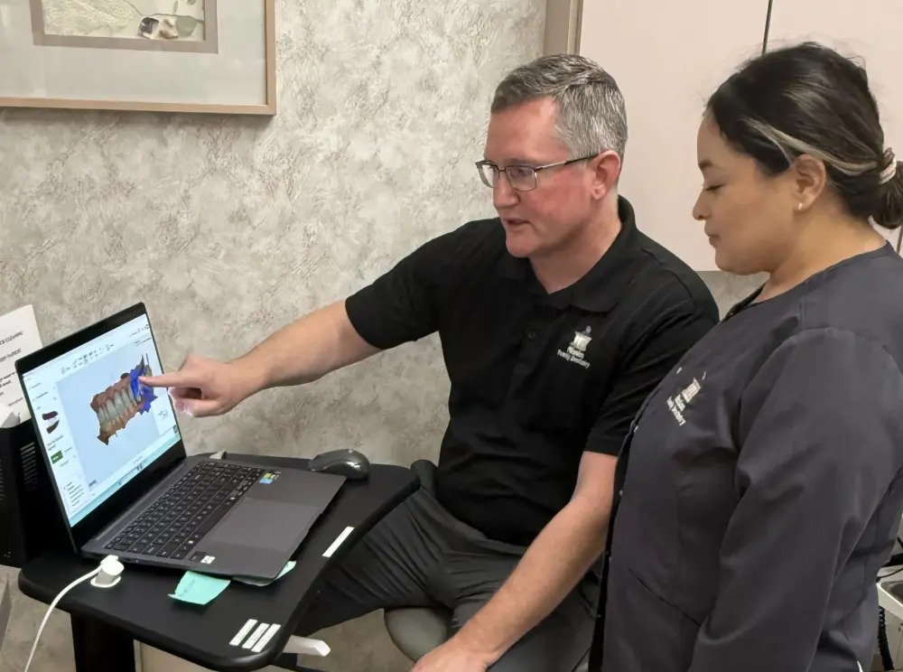
At Mission Family Dentistry, we use digital imaging technology to capture detailed views of your teeth, gums, and jaw for accurate diagnosis and treatment planning. From intraoral cameras to advanced radiographic imaging, these tools help Dr. Odenwalder identify problems early and plan care with precision.
How Digital Imaging Helps Your Dentist See More
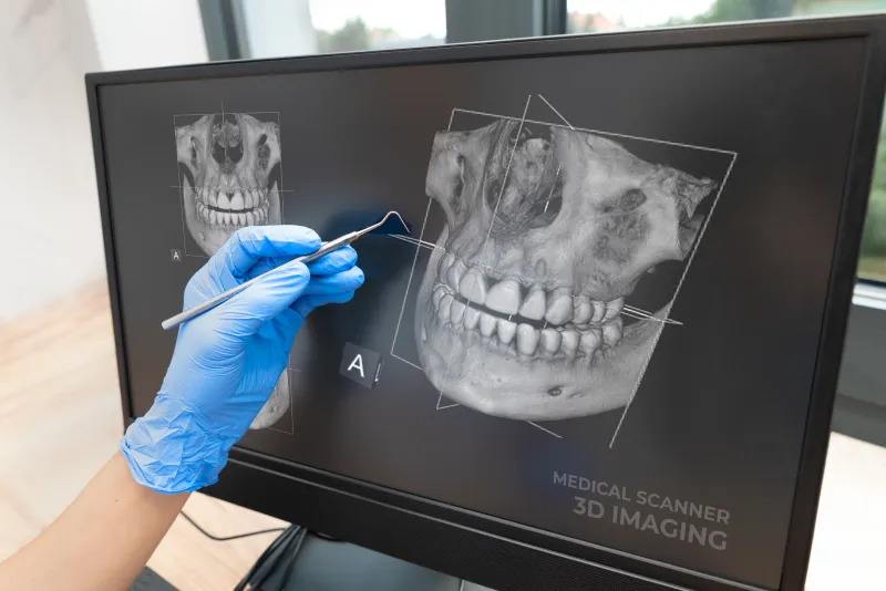
Digital dental imaging uses advanced electronic sensors and cameras to capture highly detailed views of teeth, gums, bone, and surrounding structures. Unlike older film-based methods, digital images appear on screen almost instantly and can be enhanced, magnified, and shared for more accurate diagnosis and treatment planning.
At Mission Family Dentistry in Oceanside, California, Dr. Odenwalder uses digital imaging technology to give patients a clear picture of their oral health and to plan treatments with greater precision.
Benefits of Digital Imaging
- Highly detailed images that reveal issues not visible during a standard exam.
- Instant results displayed on screen so patients can see what the dentist sees.
- Improved treatment planning for restorations, implants, and cosmetic procedures.
- Easy comparison of images over time to track changes in oral health.
- Comfortable, non-invasive process with minimal chair time.
- Secure digital storage eliminates the need for physical film archives.
Types of Digital Imaging We Use
Our office uses several forms of digital imaging depending on the clinical need:
- Intraoral cameras: A small, pen-sized camera captures close-up images of individual teeth and gum tissue. These images are displayed on a chairside monitor so you can see exactly what the dentist sees, making it easier to understand your diagnosis and treatment options.
- Digital X-rays: Electronic sensors produce detailed radiographic images with significantly less radiation than traditional film. Bitewing, periapical, and panoramic views help detect cavities, bone loss, infections, and other conditions below the surface.
- Panoramic imaging: A single, wide-format image captures the entire mouth, jaw joints, sinuses, and surrounding anatomy. This view is valuable for evaluating wisdom teeth, planning implants, and assessing overall jaw health.
What to Expect During Digital Imaging
Digital imaging is quick and comfortable. Depending on the type of image needed, a small sensor or camera is positioned in or near the mouth. The capture takes only seconds, and results appear on screen immediately for review.
There is no special preparation required. During your visit, the dental team will explain which images are being taken and why. You are welcome to view the images and ask questions about anything you see.
How Images Guide Your Care
Digital images allow Dr. Odenwalder to identify problems at the earliest stages, plan treatments with precision, and show you exactly what is happening in your mouth. Whether it is a small cavity, a crack in a restoration, or a bone-level change, imaging helps ensure nothing is missed and that your care plan is based on the most accurate information available.
Schedule Your Imaging Appointment
Want a clearer picture of your oral health? Call Mission Family Dentistry at 760-439-5515 to book an appointment with Dr. Odenwalder.
Frequently Asked Questions About Digital Imaging
Yes. Digital imaging uses significantly less radiation than traditional film methods, and intraoral cameras use no radiation at all. Protective measures such as lead aprons are used when X-rays are taken.
Most digital images are captured in seconds. A full set of images typically adds only a few minutes to your appointment.
Yes. Images appear on a chairside monitor immediately after capture. Your dentist will walk you through the findings and answer any questions.
The frequency depends on your oral health, risk factors, and treatment history. Many patients need updated images every 12 to 24 months, but your schedule is personalized to your needs.
An intraoral camera captures visible-light photos of the surfaces of teeth and gums. X-rays use a small amount of radiation to reveal structures beneath the surface, such as roots, bone, and areas between teeth.
Digital imaging is a powerful diagnostic tool, but no single method detects every condition. Your dentist combines imaging with a clinical exam, patient history, and other tests to build a complete picture of your oral health.